The vestibulocochlear nerve or auditory vestibular nerve, also known as the eighth cranial nerve, cranial nerve VIII, or simply CN VIII, is a cranial nerve that transmits sound and equilibrium (balance) information from the inner ear to the brain. Through olivocochlear fibers, it also transmits motor and modulatory information from the superior olivary complex in the brainstem to the cochlea.
>>>PUT SHARE BUTTONS HERE<<<
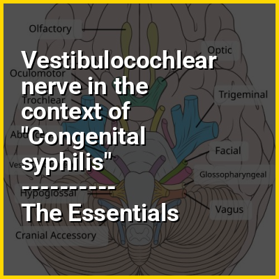
👉 Vestibulocochlear nerve in the context of Congenital syphilis
Congenital syphilis is syphilis that occurs when a mother with untreated syphilis passes the infection to her baby during pregnancy or at birth. It may present in the fetus, infant, or later. Clinical features vary and differ between early onset, that is presentation before 2-years of age, and late onset, presentation after age 2-years. Infection in the unborn baby may present as poor growth, non-immune hydrops leading to premature birth or loss of the baby, or no signs. Affected newborns mostly initially have no clinical signs. They may be small and irritable. Characteristic features include a rash, fever, large liver and spleen, a runny and congested nose, and inflammation around bone or cartilage. There may be jaundice, large glands, pneumonia (pneumonia alba), meningitis, warty bumps on genitals, deafness or blindness. Untreated babies that survive the early phase may develop skeletal deformities including deformity of the nose, lower legs, forehead, collar bone, jaw, and cheek bone. There may be a perforated or high arched palate, and recurrent joint disease. Other late signs include linear perioral tears, intellectual disability, hydrocephalus, and juvenile general paresis. Seizures and cranial nerve palsies may first occur in both early and late phases. Eighth nerve palsy, interstitial keratitis and small notched teeth may appear individually or together; known as Hutchinson's triad.
It is caused by the bacterium Treponema pallidum subspecies pallidum when it infects the baby after crossing the placenta or from contact with a syphilitic sore at birth. It is not transmitted during breastfeeding unless there is an open sore on the mother's breast. The unborn baby can become infected at any time during the pregnancy. Most cases occur due to inadequate antenatal screening and treatment during pregnancy. The baby is highly infectious if the rash and snuffles are present. The disease may be suspected from tests on the mother; blood tests and ultrasound. Tests on the baby may include blood tests, CSF analysis and medical imaging. Findings may reveal anemia and low platelets. Other findings may include low sugars, proteinuria and hypopituitarism. The placenta may appear large and pale. Other investigations include testing for HIV.
In this Dossier
- ⭐ Core Definition: Vestibulocochlear nerve
- 👉 Vestibulocochlear nerve in the context of Congenital syphilis
- Vestibulocochlear nerve in the context of Metencephalon
- Vestibulocochlear nerve in the context of Ramsay Hunt syndrome type 2
- Vestibulocochlear nerve in the context of Sensorineural hearing loss
- Vestibulocochlear nerve in the context of Auditory nerve
- Vestibulocochlear nerve in the context of Saccule
- Vestibulocochlear nerve in the context of Special somatic afferent
- Vestibulocochlear nerve in the context of Ototoxicity
Vestibulocochlear nerve in the context of Metencephalon
The metencephalon is the embryonic part of the hindbrain that differentiates into the pons and the cerebellum. It contains a portion of the fourth ventricle and the trigeminal nerve (CN V), abducens nerve (CN VI), facial nerve (CN VII), and a portion of the vestibulocochlear nerve (CN VIII).
Vestibulocochlear nerve in the context of Ramsay Hunt syndrome type 2
Ramsay Hunt syndrome type 2, commonly referred to simply as Ramsay Hunt syndrome (RHS) and also known as herpes zoster oticus, is inflammation of the geniculate ganglion of the facial nerve as a late consequence of varicella zoster virus (VZV). In regard to the frequency, less than 1% of varicella zoster infections involve the facial nerve and result in RHS. It is traditionally defined as a triad of ipsilateral facial paralysis, otalgia, and vesicles close to the ear and auditory canal. Due to its proximity to the vestibulocochlear nerve, the virus can spread and cause hearing loss, tinnitus (hearing noises that are not caused by outside sounds), and vertigo. It is common for diagnoses to be overlooked or delayed, which can raise the likelihood of long-term consequences. It is more complicated than Bell's palsy. Therapy aims to shorten its overall length, while also providing pain relief and averting any consequences.
Vestibulocochlear nerve in the context of Sensorineural hearing loss
Sensorineural hearing loss (SNHL) is a type of hearing loss in which the root cause lies in the inner ear, sensory organ (cochlea and associated structures), or the vestibulocochlear nerve (cranial nerve VIII). SNHL accounts for about 90% of reported hearing loss. SNHL is usually permanent and can be mild, moderate, severe, profound, or total. However, if the loss happened suddenly, and very recently, Prednisone and other treatments may reverse the loss (See SSHL below). Various other descriptors can be used depending on the shape of the audiogram, such as high frequency, low frequency, U-shaped, notched, peaked, or flat.
Sensory hearing loss often occurs as a consequence of damaged or deficient cochlear hair cells. Hair cells may be abnormal at birth or damaged during the lifetime of an individual. There are both external causes of damage, including infection, and ototoxic drugs, as well as intrinsic causes, including genetic mutations. A common cause or exacerbating factor in SNHL is prolonged exposure to environmental noise, or noise-induced hearing loss. Exposure to a single very loud noise, such as a gunshot or bomb blast, can cause noise-induced hearing loss. Using headphones at high volume over time, or being in loud environments regularly, such as a loud workplace, sporting events, concerts, and using noisy machines, can also be a risk for noise-induced hearing loss.
Vestibulocochlear nerve in the context of Auditory nerve
The cochlear nerve (also auditory nerve or acoustic nerve) is one of two parts of the vestibulocochlear nerve, a cranial nerve present in amniotes, the other part being the vestibular nerve. The cochlear nerve carries auditory sensory information from the cochlea of the inner ear directly to the brain. The other portion of the vestibulocochlear nerve is the vestibular nerve, which carries spatial orientation information to the brain from the semicircular canals, also known as semicircular ducts.
Vestibulocochlear nerve in the context of Saccule
The saccule (Latin: sacculus) is a bed of sensory cells in the inner ear that detects linear acceleration and head tilting in the vertical plane, and converts these vibrations into electrical impulses to be interpreted by the brain. When the head accelerates vertically, the sensory cells of the saccule are moved due to a combination of inertia and gravity. In response, the neurons connected to the saccule transmit electrical impulses that represent this movement to the brain. These impulses travel along the vestibulocochlear nerve (CNVIII) to the vestibular nuclei in the brainstem.
The vestibular system is important for balance, or equilibrium. It includes the saccule, utricle, and the three semicircular canals. The vestibule is the name of the fluid-filled, membranous duct that contains these organs of balance and is in turn encased in the temporal bone of the skull as a part of the inner ear.
Vestibulocochlear nerve in the context of Special somatic afferent
Special somatic afferent fibers (SSA) are the afferent nerve fibers that carry information from the special senses of vision, hearing and balance. The cranial nerves containing SSA fibers are the optic nerve (CN II) and the vestibulocochlear nerve (CN VIII).
Vestibulocochlear nerve in the context of Ototoxicity
Ototoxicity is the property of being toxic to the ear (oto-), specifically the cochlea or auditory nerve and sometimes the vestibular system, for example, as a side effect of a drug. The effects of ototoxicity can be reversible and temporary, or irreversible and permanent. It has been recognized since the 19th century. There are many well-known ototoxic drugs used in clinical situations, and they are prescribed, despite the risk of hearing disorders, for very serious health conditions. Ototoxic drugs include antibiotics (such as gentamicin, streptomycin, tobramycin), loop diuretics (such as furosemide), and platinum-based chemotherapy agents (such as cisplatin and carboplatin). A number of nonsteroidal anti-inflammatory drugs (NSAIDS) have also been shown to be ototoxic. This can result in sensorineural hearing loss, dysequilibrium, or both. Some environmental and occupational chemicals have also been shown to affect the auditory system and interact with noise.