Myofilament in the context of "Myofibril"

Play Trivia Questions online!

or

Skip to study material about Myofilament in the context of "Myofibril"

Ad spacer

>>>PUT SHARE BUTTONS HERE<<<

👉 Myofilament in the context of Myofibril

A myofibril (also known as a muscle fibril or sarcostyle) is a basic rod-like organelle of a muscle cell. Skeletal muscles are composed of long, tubular cells known as muscle fibers, and these cells contain many chains of myofibrils. Each myofibril has a diameter of 1–2 micrometres. They are created during embryonic development in a process known as myogenesis.

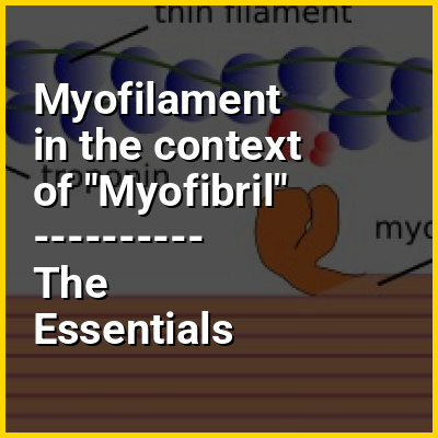
Myofibrils are composed of long proteins including actin, myosin, and titin, and other proteins that hold them together. These proteins are organized into thick, thin, and elastic myofilaments, which repeat along the length of the myofibril in sections or units of contraction called sarcomeres. Muscles contract by sliding the thick myosin, and thin actin myofilaments along each other.

↓ Explore More Topics
In this Dossier

Myofilament in the context of Muscular contraction

Muscle contraction is the activation of tension-generating sites within muscle cells. In physiology, muscle contraction does not necessarily mean muscle shortening because muscle tension can be produced without changes in muscle length (isometric contraction), such as when holding something heavy in the same position. The termination of muscle contraction is followed by muscle relaxation, which is a return of the muscle fibers to their low tension-generating state.

For the contractions to happen, the muscle cells must rely on the change in action of two types of filament: thin and thick filaments.

↑ Return to Menu

Myofilament in the context of Myocardium

Cardiac muscle (also called heart muscle or myocardium) is one of three types of vertebrate muscle tissues, the others being skeletal muscle and smooth muscle. It is an involuntary, striated muscle that constitutes the main tissue of the wall of the heart. The cardiac muscle (myocardium) forms a thick middle layer between the outer layer of the heart wall (the pericardium) and the inner layer (the endocardium), with blood supplied via the coronary circulation. It is composed of individual cardiac muscle cells joined by intercalated discs, and encased by collagen fibers and other substances that form the extracellular matrix.

Cardiac muscle contracts in a similar manner to skeletal muscle, although with some important differences. Electrical stimulation in the form of a cardiac action potential triggers the release of calcium from the cell's internal calcium store, the sarcoplasmic reticulum. The rise in calcium causes the cell's myofilaments to slide past each other in a process called excitation-contraction coupling.Diseases of the heart muscle known as cardiomyopathies are of major importance. These include ischemic conditions caused by a restricted blood supply to the muscle such as angina, and myocardial infarction.

↑ Return to Menu