A motor nerve, or efferent nerve, is a nerve that contains exclusively efferent nerve fibers and transmits motor signals from the central nervous system (CNS) to the effector organs (muscles and glands), as opposed to sensory nerves, which transfer signals from sensory receptors in the periphery to the CNS. This is different from the motor neuron, which includes a cell body and branching of dendrites, while the nerve is made up of a bundle of axons. In the strict sense, a "motor nerve" can refer exclusively to the connection to muscles, excluding other organs. The vast majority of nerves contain both sensory and motor fibers and are therefore called mixed nerves.
>>>PUT SHARE BUTTONS HERE<<<
👉 Motor nerve in the context of Sensory nerve
A sensory nerve, or afferent nerve, is a nerve that contains exclusively afferent nerve fibers. Nerves containing also motor fibers are called mixed. Afferent nerve fibers in a sensory nerve carry sensory information toward the central nervous system (CNS) from different sensory receptors of sensory neurons in the peripheral nervous system (PNS).
Contrarily, a motor nerve carries information from the CNS to the PNS.
In this Dossier
Motor nerve in the context of Nervous system
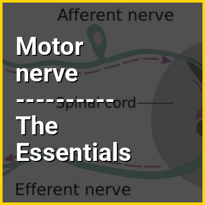
In biology, the nervous system is the highly complex part of an animal that coordinates its actions and sensory information by transmitting signals to and from different parts of its body. The nervous system detects environmental changes that impact the body, then works in tandem with the endocrine system to respond to such events. Nervous tissue first arose in wormlike organisms about 550 to 600 million years ago. In vertebrates, it consists of two main parts, the central nervous system (CNS) and the peripheral nervous system (PNS). The CNS consists of the brain and spinal cord. The PNS consists mainly of nerves, which are enclosed bundles of the long fibers, or axons, that connect the CNS to every other part of the body. Nerves that transmit signals from the brain are called motor nerves (efferent), while those nerves that transmit information from the body to the CNS are called sensory nerves (afferent). The PNS is divided into two separate subsystems, the somatic and autonomic nervous systems. The autonomic nervous system is further subdivided into the sympathetic, parasympathetic and enteric nervous systems. The sympathetic nervous system is activated in cases of emergencies to mobilize energy, while the parasympathetic nervous system is activated when organisms are in a relaxed state. The enteric nervous system functions to control the gastrointestinal system. Nerves that exit from the brain are called cranial nerves while those exiting from the spinal cord are called spinal nerves.
The nervous system consists of nervous tissue which, at a cellular level, is defined by the presence of a special type of cell, called the neuron. Neurons have special structures that allow them to send signals rapidly and precisely to other cells. They send these signals in the form of electrochemical impulses traveling along thin fibers called axons, which can be directly transmitted to neighboring cells through electrical synapses or cause chemicals called neurotransmitters to be released at chemical synapses. A cell that receives a synaptic signal from a neuron may be excited, inhibited, or otherwise modulated. The connections between neurons can form neural pathways, neural circuits, and larger networks that generate an organism's perception of the world and determine its behavior. Along with neurons, the nervous system contains other specialized cells called glial cells (or simply glia), which provide structural and metabolic support. Many of the cells and vasculature channels within the nervous system make up the neurovascular unit, which regulates cerebral blood flow in order to rapidly satisfy the high energy demands of activated neurons.
Motor nerve in the context of Neuropathy
Peripheral neuropathy, often shortened to neuropathy, refers to damage or disease affecting the nerves. Damage to nerves may impair sensation, movement, gland function, and/or organ function depending on which nerve fibers are affected. Neuropathies affecting motor, sensory, or autonomic nerve fibers result in different symptoms. More than one type of fiber may be affected simultaneously. Peripheral neuropathy may be acute (with sudden onset, rapid progress) or chronic (symptoms begin subtly and progress slowly), and may be reversible or permanent.
Common causes include systemic diseases (such as diabetes or leprosy), hyperglycemia-induced glycation, vitamin deficiency, medication (e.g., chemotherapy, or commonly prescribed antibiotics including metronidazole and the fluoroquinolone class of antibiotics (such as ciprofloxacin, levofloxacin, moxifloxacin)), traumatic injury, ischemia, radiation therapy, excessive alcohol consumption, immune system disease, celiac disease, non-celiac gluten sensitivity, or viral infection. It can also be genetic (present from birth) or idiopathic (no known cause). In conventional medical usage, the word neuropathy (neuro-, "nervous system" and -pathy, "disease of") without modifier usually means peripheral neuropathy.
Motor nerve in the context of Secretomotor
The adjective secretomotor refers to the capacity of a structure (often a nerve) to induce a gland to secrete a substance (usually mucus or serous fluid).
Secretomotor nerve endings are frequently contrasted with sensory neuron endings and motor nerve endings. An example of secretomotor activity can be seen with the lacrimal gland, which secretes the aqueous layer of the tear film. The lacrimal branch of the ophthalmic nerve (itself a branch of trigeminal nerve V1) supplies secretomotor innervation to the lacrimal gland, stimulating its secretion of the aqueous layer. However, these nerves fibers originate from the facial nerve (VII) and only travel briefly with fibers from the trigeminal nerve.
Motor nerve in the context of Nerve conduction study
A nerve conduction study (NCS) is a medical diagnostic test commonly used to evaluate the function, especially the ability of electrical conduction, of the motor and sensory nerves of the human body. These tests may be performed by medical specialists such as clinical neurophysiologists, physical therapists, physiatrists (physical medicine and rehabilitation physicians), and neurologists who subspecialize in electrodiagnostic medicine. In the United States, neurologists and physiatrists receive training in electrodiagnostic medicine (performing needle electromyography (EMG) and NCSs) as part of residency training and, in some cases, acquire additional expertise during a fellowship in clinical neurophysiology, electrodiagnostic medicine, or neuromuscular medicine. Outside the US, clinical neurophysiologists learn needle EMG and NCS testing.