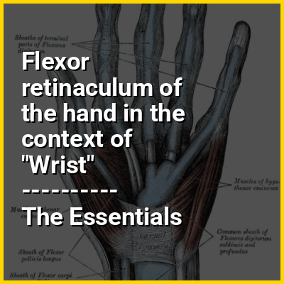
The flexor retinaculum (transverse carpal ligament or anterior annular ligament) is a fibrous band on the palmar side of the hand near the wrist. It arches over the carpal bones of the hands, covering them and forming the carpal tunnel.
>>>PUT SHARE BUTTONS HERE<<<
👉 Flexor retinaculum of the hand in the context of Wrist
In human anatomy, the wrist is variously defined as (1) the carpus or carpal bones, the complex of eight bones forming the proximal skeletal segment of the hand; (2) the wrist joint or radiocarpal joint, the joint between the radius and the carpus and; (3) the anatomical region surrounding the carpus including the distal parts of the bones of the forearm and the proximal parts of the metacarpus or five metacarpal bones and the series of joints between these bones, thus referred to as wrist joints. This region also includes the carpal tunnel, the anatomical snuff box, bracelet lines, the flexor retinaculum, and the extensor retinaculum.
As a consequence of these various definitions, fractures to the carpal bones are referred to as carpal fractures, while fractures such as distal radius fracture are often considered fractures to the wrist.
In this Dossier
Flexor retinaculum of the hand in the context of Carpal tunnel
In the human body, the carpal tunnel or carpal canal is a flattened body cavity on the flexor (palmar/volar) side of the wrist, bounded by the carpal bones and flexor retinaculum. It forms the passageway that transmits the median nerve and the tendons of the extrinsic flexor muscles of the hand from the forearm to the hand. The median artery is an anatomical variant (increasingly found). When present it lies between the radial artery, and the ulnar artery and runs with the median nerve supplying the same structures innervated.
When swelling or degeneration occurs in the tendons and sheaths of any of the nine flexor muscles (flexor pollicis longus, four flexor digitorum profundus and four flexor digitorum superficialis) passing through the carpal tunnel, the canal can narrow and compress/entrap the median nerve, resulting in a compression neuropathy known as carpal tunnel syndrome (CTS). If untreated, neuropraxia, parasthesia and muscle atrophy (especially of the thenar muscles) can occur. The condition often requires surgical division of the retinaculum to relieve the pressure upon the nerve.