Diencephalon in the context of "Brain"

⭐ In the context of the brain, the diencephalon is considered a subdivision of which larger embryonic brain region?

Ad spacer

⭐ Core Definition: Diencephalon

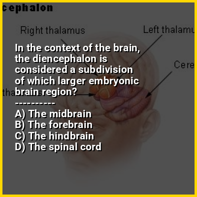
In the human brain, the diencephalon (or interbrain) is a division of the forebrain (embryonic prosencephalon). It is situated between the telencephalon and the midbrain (embryonic mesencephalon). The diencephalon has also been known as the tweenbrain in older literature. It consists of structures that are on either side of the third ventricle, including the thalamus, the hypothalamus, the epithalamus and the subthalamus.

The diencephalon is one of the main vesicles of the brain formed during embryonic development. During the third week of development a neural tube is created from the ectoderm, one of the three primary germ layers, and forms three main vesicles: the prosencephalon, the mesencephalon and the rhombencephalon. The prosencephalon gradually divides into the telencephalon (the cerebrum) and the diencephalon.

↓ Menu

>>>PUT SHARE BUTTONS HERE<<<

👉 Diencephalon in the context of Brain

The brain is an organ that serves as the center of the nervous system in all vertebrate and most invertebrate animals. It consists of nervous tissue and is typically located in the head (cephalization), usually near organs for special senses such as vision, hearing, and olfaction. Being the most specialized organ, it is responsible for receiving information from the sensory nervous system, processing that information (thought, cognition, and intelligence) and the coordination of motor control (muscle activity and endocrine system).

While invertebrate brains arise from paired segmental ganglia (each of which is only responsible for the respective body segment) of the ventral nerve cord, vertebrate brains develop axially from the midline dorsal nerve cord as a vesicular enlargement at the rostral end of the neural tube, with centralized control over all body segments. All vertebrate brains can be embryonically divided into three parts: the forebrain (prosencephalon, subdivided into telencephalon and diencephalon), midbrain (mesencephalon) and hindbrain (rhombencephalon, subdivided into metencephalon and myelencephalon). The spinal cord, which directly interacts with somatic functions below the head, can be considered a caudal extension of the myelencephalon enclosed inside the vertebral column. Together, the brain and spinal cord constitute the central nervous system in all vertebrates.

↓ Explore More Topics
In this Dossier

Diencephalon in the context of Brainstem

The brainstem (or brain stem) is the posterior stalk-like part of the brain that connects the cerebrum with the spinal cord. In the human brain the brainstem is composed of the midbrain, the pons, and the medulla oblongata. The midbrain is continuous with the thalamus of the diencephalon through the tentorial notch, and sometimes the diencephalon is included in the brainstem.

The brainstem is very small, making up around only 2.6 percent of the brain's total weight. It has the critical roles of regulating heart and respiratory function, helping to control heart rate and breathing rate. It also provides the main motor and sensory nerve supply to the face and neck via the cranial nerves. Ten pairs of cranial nerves come from the brainstem. Other roles include the regulation of the central nervous system and the body's sleep cycle. It is also of prime importance in the conveyance of motor and sensory pathways from the rest of the brain to the body, and from the body back to the brain. These pathways include the corticospinal tract (motor function), the dorsal column-medial lemniscus pathway (fine touch, vibration sensation, and proprioception), and the spinothalamic tract (pain, temperature, itch, and crude touch).

↑ Return to Menu

Diencephalon in the context of Peripheral nervous system

The peripheral nervous system (PNS) is one of two components that make up the nervous system of bilateral animals, with the other part being the central nervous system (CNS). The PNS consists of nerves and ganglia, which lie outside the brain and the spinal cord. The main function of the PNS is to connect the CNS to the limbs and organs, essentially serving as a relay between the brain and spinal cord and the rest of the body. Unlike the CNS, the PNS is not protected by the vertebral column and skull, or by the blood–brain barrier, which leaves it exposed to toxins.

The peripheral nervous system can be divided into a somatic division and an autonomic division. Each of these can further be differentiated into a sensory and a motor sector. In the somatic nervous system, the cranial nerves are part of the PNS with the exceptions of the olfactory nerve and epithelia and the optic nerve (cranial nerve II) along with the retina, which are considered parts of the central nervous system based on developmental origin. The second cranial nerve is not a true peripheral nerve but a tract of the diencephalon. Cranial nerve ganglia, as with all ganglia, are part of the PNS. The autonomic nervous system exerts involuntary control over smooth muscle and glands.

↑ Return to Menu

Diencephalon in the context of Forebrain

In the anatomy of the brain of vertebrates, the forebrain or prosencephalon is the rostral (forward-most) portion of the brain. The forebrain controls body temperature, reproductive functions, eating, sleeping, and the display of emotions.

Vesicles of the forebrain (prosencephalon), the midbrain (mesencephalon), and hindbrain (rhombencephalon) are the three primary brain vesicles during the early development of the nervous system. At the five-vesicle stage, the forebrain separates into the diencephalon (thalamus, hypothalamus, subthalamus, and epithalamus) and the telencephalon which develops into the cerebrum. The cerebrum consists of the cerebral cortex, underlying white matter, and the basal ganglia.

↑ Return to Menu

Diencephalon in the context of Midbrain

The midbrain or mesencephalon is the uppermost portion of the brainstem connecting the diencephalon and cerebrum with the pons. It consists of the cerebral peduncles, tegmentum, and tectum.

It is functionally associated with vision, hearing, motor control, sleep and wakefulness, arousal (alertness), and temperature regulation.

↑ Return to Menu

Diencephalon in the context of Thalamus

The thalamus (pl.: thalami; from Greek θάλαμος, "chamber") is a large mass of gray matter on the lateral wall of the third ventricle forming the dorsal part of the diencephalon (a division of the forebrain). Nerve fibers project out of the thalamus to the cerebral cortex in all directions, known as the thalamocortical radiations, allowing hub-like exchanges of information. It has several functions, such as the relaying of sensory and motor signals to the cerebral cortex and the regulation of consciousness, sleep, and alertness.

Anatomically, the thalami are paramedian symmetrical structures (left and right), within the vertebrate brain, situated between the cerebral cortex and the midbrain. It forms during embryonic development as the main product of the diencephalon, as first recognized by the Swiss embryologist and anatomist Wilhelm His Sr. in 1893.

↑ Return to Menu

Diencephalon in the context of Tentorial notch

The tentorial notch (also known as the tentorial incisure or incisura tentorii) refers to the anterior opening between the free edge of the cerebellar tentorium and the clivus for the passage of the brainstem.

The midbrain continues with the thalamus of the diencephalon through the tentorial notch.

↑ Return to Menu

Diencephalon in the context of Fornix (neuroanatomy)

The fornix (from Latin: fornix, lit.'arch'; pl.: fornices) is a C-shaped bundle of nerve fibers in the brain that acts as the major output tract of the hippocampus. The fornix also carries some afferent fibers to the hippocampus from structures in the diencephalon and basal forebrain. The fornix is part of the limbic system. While its exact function and importance in the physiology of the brain are still not entirely clear, it has been demonstrated in humans that surgical transection—the cutting of the fornix along its body—can cause memory loss. There is some debate over what type of memory is affected by this damage, but it has been found to most closely correlate with recall memory rather than recognition memory. This means that damage to the fornix can cause difficulty in recalling long-term information such as details of past events, but it has little effect on the ability to recognize objects or familiar situations.

↑ Return to Menu