Thoracic outlet syndrome in the context of Nerve conduction studies


Thoracic outlet syndrome in the context of Nerve conduction studies

⭐ Core Definition: Thoracic outlet syndrome

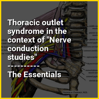
Thoracic outlet syndrome (TOS) is a condition in which there is compression of the nerves, arteries, or veins in the superior thoracic aperture, the passageway from the lower neck to the armpit, also known as the thoracic outlet. There are three main types: neurogenic, venous, and arterial. The neurogenic type is the most common and presents with pain, weakness, paraesthesia, and occasionally loss of muscle at the base of the thumb. The venous type results in swelling, pain, and possibly a bluish coloration of the arm. The arterial type results in pain, coldness, and pallor of the arm.

TOS may result from trauma, repetitive arm movements, tumors, pregnancy, or anatomical variations such as a cervical rib. The diagnosis may be supported by nerve conduction studies and medical imaging. TOS is difficult to diagnose and there are many potential differential diagnoses as well as other diseases that are often co-occurrent with TOS.

↓ Menu
HINT:

In this Dossier

Thoracic outlet syndrome in the context of Cervical rib

Cervical ribs are the ribs of the neck in many tetrapods. In most mammals, including humans, cervical ribs are not normally present as separate structures. They can, however, occur as a pathology. In humans, pathological cervical ribs are usually not of clinical concern, although they can cause a form of thoracic outlet syndrome.

View the full Wikipedia page for Cervical rib
↑ Return to Menu

Thoracic outlet syndrome in the context of Thoracic inlet

The superior thoracic aperture, also known as the thoracic outlet, or thoracic inlet refers to the opening at the top of the thoracic cavity. It is also clinically referred to as the thoracic outlet, in the case of thoracic outlet syndrome. A lower thoracic opening is the inferior thoracic aperture.

View the full Wikipedia page for Thoracic inlet
↑ Return to Menu

Thoracic outlet syndrome in the context of Magnetic resonance neurography

Magnetic resonance neurography (MRN) is the direct imaging of nerves in the body by optimizing selectivity for unique MRI water properties of nerves. It is a modification of magnetic resonance imaging. This technique yields a detailed image of a nerve from the resonance signal that arises from in the nerve itself rather than from surrounding tissues or from fat in the nerve lining. Because of the intraneural source of the image signal, the image provides a medically useful set of information about the internal state of the nerve such as the presence of irritation, nerve swelling (edema), compression, pinch or injury. Standard magnetic resonance images can show the outline of some nerves in portions of their courses but do not show the intrinsic signal from nerve water. Magnetic resonance neurography is used to evaluate major nerve compressions such as those affecting the sciatic nerve (e.g. piriformis syndrome), the brachial plexus nerves (e.g. thoracic outlet syndrome), the pudendal nerve, or virtually any named nerve in the body. A related technique for imaging neural tracts in the brain and spinal cord is called magnetic resonance tractography or diffusion tensor imaging.

View the full Wikipedia page for Magnetic resonance neurography
↑ Return to Menu