Endometrium in the context of "Pap test"

Play Trivia Questions online!

or

Skip to study material about Endometrium in the context of "Pap test"

Ad spacer

>>>PUT SHARE BUTTONS HERE<<<
In this Dossier

Endometrium in the context of Uterus

The uterus (from Latin uterus, pl.: uteri or uteruses) or womb (/wm/) is the organ in the reproductive system of most female mammals, including humans, that accommodates the embryonic and fetal development of one or more fertilized eggs until birth. The uterus is a hormone-responsive sex organ that contains glands in its lining that secrete uterine milk for embryonic nourishment. (The term uterus is also applied to analogous structures in some non-mammalian animals.)

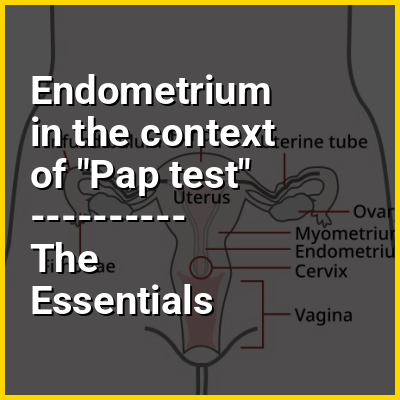
In humans, the lower end of the uterus is a narrow part known as the isthmus that connects to the cervix, the anterior gateway leading to the vagina. The upper end, the body of the uterus, is connected to the fallopian tubes at the uterine horns; the rounded part, the fundus, is above the openings to the fallopian tubes. The connection of the uterine cavity with a fallopian tube is called the uterotubal junction. The fertilized egg is carried to the uterus along the fallopian tube. It will have divided on its journey to form a blastocyst that will implant itself into the lining of the uterus – the endometrium, where it will receive nutrients and develop into the embryo proper, and later fetus, for the duration of the pregnancy.

↑ Return to Menu

Endometrium in the context of Menstruation

Menstruation (also known as a period, among other colloquial terms) is the regular discharge of blood and mucosal tissue from the inner lining of the uterus through the vagina. The menstrual cycle is characterized by the rise and fall of hormones. Menstruation is triggered by falling progesterone levels, and is a sign that pregnancy has not occurred. Feminine hygiene products are used in order to maintain hygiene during menses.

The first period, a point in time known as menarche, usually begins during puberty, between the ages of 11 and 13. However, menstruation starting as young as eight years would still be considered normal. The average age of the first period is generally later in the developing world, and earlier in the developed world. The typical length of time between the first day of one period and the first day of the next is 21 to 45 days in young women; in adults, the range is between 21 and 35 days with the average often cited as 28 days. In the largest study of menstrual app data, the mean menstrual cycle length was determined to be 29.3 days. Bleeding typically lasts two to seven days. Periods stop during pregnancy and typically do not resume during the initial months of breastfeeding. Lochia occurs after childbirth. Menstruation, and with it the possibility of pregnancy, ceases after menopause, which usually occurs between 45 and 55 years of age.

↑ Return to Menu

Endometrium in the context of Implantation (embryology)

Implantation, also known as nidation, is the stage in the mammalian embryonic development in which the blastocyst hatches, attaches, adheres, and invades into the endometrium of the female's uterus. Implantation is the first stage of gestation, and, when successful, the female is considered to be pregnant. An implanted embryo is detected by the presence of increased levels of human chorionic gonadotropin (hCG) in a pregnancy test. The implanted embryo will receive oxygen and nutrients in order to grow.

For implantation to take place the uterus must become receptive. Uterine receptivity involves much cross-talk between the embryo and the uterus, initiating changes to the endometrium. This stage gives a synchrony that opens a window of implantation that enables successful implantation of a viable embryo. The endocannabinoid system plays a vital role in this synchrony in the uterus, influencing uterine receptivity, and embryo implantation. The embryo expresses cannabinoid receptors early in its development that are responsive to anandamide (AEA) secreted in the uterus. AEA is produced at higher levels before implantation and is then down-regulated at the time of implantation. This signaling is of importance in the embryo-uterus crosstalk in regulating the timing of embryonic implantation and uterine receptivity. Adequate concentrations of AEA that are neither too high or too low, are needed for successful implantation.

↑ Return to Menu

Endometrium in the context of Placenta

The placenta (pl.: placentas or placentae) is a temporary embryonic and later fetal organ that begins developing from the blastocyst shortly after implantation. It plays critical roles in facilitating nutrient, gas, and waste exchange between the physically separate maternal and fetal circulations, and is an important endocrine organ, producing hormones that regulate both maternal and fetal physiology during pregnancy. The placenta connects to the fetus via the umbilical cord, and on the opposite aspect to the maternal uterus in a species-dependent manner. In humans, a thin layer of maternal decidual (endometrial) tissue comes away with the placenta when it is expelled from the uterus following birth (sometimes incorrectly referred to as the 'maternal part' of the placenta). Placentas are a defining characteristic of placental mammals, but are also found in marsupials and some non-mammals with varying levels of development.

Mammalian placentas probably first evolved about 150 million to 200 million years ago. The protein syncytin, found in the outer barrier of the placenta (the syncytiotrophoblast) between mother and fetus, has a certain RNA signature in its genome that has led to the hypothesis that it originated from an ancient retrovirus: essentially a virus that helped pave the transition from egg-laying to live-birth.

↑ Return to Menu

Endometrium in the context of Female reproductive tract

The human female reproductive system is made up of the internal and external sex organs that function in the reproduction of new offspring. The reproductive system is immature at birth and develops at puberty to be able to release matured ova from the ovaries, facilitate their fertilization, and create a protective environment for the developing fetus during pregnancy. The female reproductive tract is made of several connected internal sex organs—the vagina, uterus, and fallopian tubes—and is prone to infections. The vagina allows for sexual intercourse and childbirth, and is connected to the uterus at the cervix. The uterus (or womb) accommodates the embryo by developing the uterine lining.

The uterus also produces secretions which help the transit of sperm to the fallopian tubes, where sperm fertilize the ova. During the menstrual cycle, the ovaries release an ovum, which transits through the fallopian tube into the uterus. If an egg cell meets with sperm on its way to the uterus, a single sperm cell can enter and merge with it, creating a zygote. If no fertilization occurs, menstruation is the process by which the uterine lining is shed as blood, mucus, and tissue.

↑ Return to Menu

Endometrium in the context of Decidua

The decidua is the modified mucosal lining of the uterus (that is, modified endometrium) that forms every month, in preparation for pregnancy. It is shed off each month when there is no fertilized egg to support. The decidua is under the influence of progesterone. Endometrial cells become highly characteristic. The decidua forms the maternal part of the placenta and remains for the duration of the pregnancy. After birth the decidua is shed together with the placenta.

↑ Return to Menu

Endometrium in the context of Blastocyst

The blastocyst is a structure formed in the early embryonic development of mammals. It possesses an inner cell mass (ICM) also known as the embryoblast which subsequently forms the embryo, and an outer layer of trophoblast cells called the trophectoderm. This layer surrounds the inner cell mass and a fluid-filled cavity or lumen known as the blastocoel. In the late blastocyst, the trophectoderm is known as the trophoblast. The trophoblast gives rise to the chorion and amnion, the two fetal membranes that surround the embryo. The placenta derives from the embryonic chorion (the portion of the chorion that develops villi). The corresponding structure in non-mammalian animals is an undifferentiated ball of cells called the blastula.

In humans, blastocyst formation begins about five days after fertilization when a fluid-filled cavity opens up in the morula, the early embryonic stage of a ball of 16 cells.The blastocyst has a diameter of about 0.1–0.2 mm and comprises 100-200 cells following 7-8 rounds of cleavage (cell division without cell growth). About seven days after fertilization, the blastocyst undergoes implantation, embedding into the endometrium of the uterine wall where it will undergo further developmental processes, including gastrulation. Embedding of the blastocyst into the endometrium requires that it hatches from the zona pellucida, the egg coat that prevents adherence to the fallopian tube as the pre-embryo makes its way to the uterus.

↑ Return to Menu