Earlobe in the context of "Outer ear"

Play Trivia Questions online!

or

Skip to study material about Earlobe in the context of "Outer ear"

Ad spacer

⭐ Core Definition: Earlobe

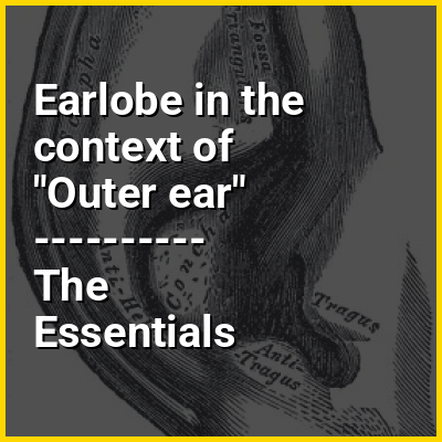
The human earlobe (lobulus auriculae), the lower portion of the outer ear, is composed of tough areolar and adipose connective tissues, lacking the firmness and elasticity of the rest of the auricle (the external structure of the ear). In some cases the lower lobe is connected to the side of the face. Since the earlobe does not contain cartilage it has a large blood supply and may help to warm the ears. However, earlobes are not generally considered to have any major biological function. The earlobe contains many nerve endings, and for some people is an erogenous zone.

The zoologist Desmond Morris in his book The Naked Ape (1967) conjectured that the lobes developed as an additional erogenous zone to facilitate the extended sexuality necessary in the evolution of human monogamous pair bonding.

↓ Menu

>>>PUT SHARE BUTTONS HERE<<<
In this Dossier

Earlobe in the context of Pulse oximetry

Pulse oximetry is a noninvasive method for monitoring blood oxygen saturation. Peripheral oxygen saturation (SpO2) readings are typically within 2% accuracy (within 4% accuracy in 95% of cases) of the more accurate (and invasive) reading of arterial oxygen saturation (SaO2) from arterial blood gas analysis.

A standard pulse oximeter passes two wavelengths of light through tissue to a photodetector. Taking advantage of the pulsate flow of arterial blood, it measures the change in absorbance over the course of a cardiac cycle, allowing it to determine the absorbance due to arterial blood alone, excluding unchanging absorbance due to venous blood, skin, bone, muscle, fat, and, in many cases, nail polish. The two wavelengths measure the quantities of bound (oxygenated) and unbound (non-oxygenated) hemoglobin, and from their ratio, the percentage of bound hemoglobin is computed.The most common approach is transmissive pulse oximetry. In this approach, one side of a thin part of the patient's body, usually a fingertip or earlobe, is illuminated, and the photodetector is on the other side. Fingertips and earlobes have disproportionately high blood flow relative to their size, in order to keep warm, but this will be lacking in hypothermic patients. Other convenient sites include an infant's foot or an unconscious patient's cheek or tongue.

↑ Return to Menu