Cone cell in the context of "Optic disc"

Play Trivia Questions online!

or

Skip to study material about Cone cell in the context of "Optic disc"

Ad spacer

⭐ Core Definition: Cone cell

Cone cells or cones are photoreceptor cells in the retina of the vertebrate eye. Cones are active in daylight conditions and enable photopic vision, as opposed to rod cells, which are active in dim light and enable scotopic vision. Most vertebrates (including humans) have several classes of cones, each sensitive to a different part of the visible spectrum of light. The comparison of the responses of different cone cell classes enables color vision. There are about six to seven million cones in a human eye (vs ~92 million rods), with the highest concentration occurring towards the macula and most densely packed in the fovea centralis, a 0.3 mm diameter rod-free area with very thin, densely packed cones. Conversely, like rods, they are absent from the optic disc, contributing to the blind spot.

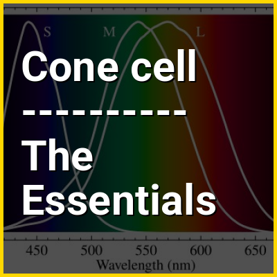
Cones are less sensitive to light than the rod cells in the retina (which support vision at low light levels), but allow the perception of color. They are also able to perceive finer detail and more rapid changes in images because their response times to stimuli are faster than those of rods. In humans, cones are normally one of three types: S-cones, M-cones and L-cones, with each type bearing a different opsin: OPN1SW, OPN1MW, and OPN1LW respectively. These cones are sensitive to visible wavelengths of light that correspond to short-wavelength, medium-wavelength and longer-wavelength light respectively. Because humans usually have three kinds of cones with different photopsins, which have different response curves and thus respond to variation in color in different ways, humans have trichromatic vision. Being color blind can change this, and there have been some verified reports of people with four types of cones, giving them tetrachromatic vision.The three pigments responsible for detecting light have been shown to vary in their exact chemical composition due to genetic mutation; different individuals will have cones with different color sensitivity.

↓ Menu

>>>PUT SHARE BUTTONS HERE<<<
In this Dossier

Cone cell in the context of Color

Color (or colour in Commonwealth English) is the visual perception produced by the activation of the different types of cone cells in the eye caused by light. Though color is not an inherent property of matter, color perception is related to an object's light absorption, emission, reflection and transmission. For most humans, visible wavelengths of light are the ones perceived in the visible light spectrum, with three types of cone cells (trichromacy). Other animals may have a different number of cone cell types or have eyes sensitive to different wavelengths, such as bees that can distinguish ultraviolet, and thus have a different color sensitivity range. Animal perception of color originates from different light wavelength or spectral sensitivity in cone cell types, which is then processed by the brain.

Colors have perceived properties such as hue, colorfulness, and lightness. Colors can also be additively mixed (mixing light) or subtractively mixed (mixing pigments). If one color is mixed in the right proportions, because of metamerism, they may look the same as another stimulus with a different reflection or emission spectrum. For convenience, colors can be organized in a color space, which when being abstracted as a mathematical color model can assign each region of color with a corresponding set of numbers. As such, color spaces are an essential tool for color reproduction in print, photography, computer monitors, and television. Some of the most well-known color models and color spaces are RGB, CMYK, HSL/HSV, CIE Lab, and YCbCr/YUV.

↑ Return to Menu

Cone cell in the context of Sensory nervous system

The sensory nervous system is a part of the nervous system responsible for processing sensory information. A sensory system consists of sensory neurons (including the sensory receptor cells), neural pathways, and parts of the brain involved in sensory perception and interoception. Commonly recognized sensory systems are those for vision, hearing, touch, taste, smell, balance and visceral sensation. Sense organs are transducers that convert data from the outer physical world to the realm of the mind where people interpret the information, creating their perception of the world around them.

The receptive field is the area of the body or environment to which a receptor organ and receptor cells respond. For instance, the part of the world an eye can see, is its receptive field; the light that each rod or cone can see, is its receptive field. Receptive fields have been identified for the visual system, auditory system and somatosensory system.

↑ Return to Menu

Cone cell in the context of Trichromacy

Trichromacy or trichromatism is the possession of three independent channels for conveying color information, derived from the three different types of cone cells in the eye. Organisms with trichromacy are called trichromats.

The normal explanation of trichromacy is that the organism's retina contains three types of color receptors (called cone cells in vertebrates) with different absorption spectra. In actuality, the number of such receptor types may be greater than three, since different types may be active at different light intensities. In vertebrates with three types of cone cells, at low light intensities the rod cells may contribute to color vision.

↑ Return to Menu

Cone cell in the context of Spectral sensitivity

Spectral sensitivity is the relative efficiency of detection, of light or other signal, as a function of the frequency or wavelength of the signal.

In visual neuroscience, spectral sensitivity is used to describe the different characteristics of the photopigments in the rod cells and cone cells in the retina of the eye. It is known that the rod cells are more suited to scotopic vision and cone cells to photopic vision, and that they differ in their sensitivity to different wavelengths of light. It has been established that the maximum spectral sensitivity of the human eye under daylight conditions is at a wavelength of 555 nm, while at night the peak shifts to 507 nm.

↑ Return to Menu

Cone cell in the context of Metamerism (color)

In colorimetry, metamerism is a perceived matching of colors with different (nonmatching) spectral power distributions. Colors that match this way are called metamers.

A spectral power distribution describes the proportion of total light given off (emitted, transmitted, or reflected) by a color sample at each visible wavelength; it defines the complete information about the light coming from the sample. However, the human eye contains only three color receptors (three types of cone cells), which means that all colors are reduced to three sensory quantities, called the tristimulus values. Metamerism occurs because each type of cone responds to the cumulative energy from a broad range of wavelengths, so that different combinations of light across all wavelengths can produce an equivalent receptor response and the same tristimulus values or color sensation. In color science, the set of sensory spectral sensitivity curves is numerically represented by color matching functions.

↑ Return to Menu

Cone cell in the context of Retina

The retina (from Latin rete 'net'; pl.retinae or retinas) is the innermost, light-sensitive layer of tissue of the eye of most vertebrates and some molluscs. The optics of the eye create a focused two-dimensional image of the visual world on the retina, which then processes that image within the retina and sends nerve impulses along the optic nerve to the visual cortex to create visual perception. The retina serves a function which is in many ways analogous to that of the film or image sensor in a camera.

The neural retina consists of several layers of neurons interconnected by synapses and is supported by an outer layer of pigmented epithelial cells. The primary light-sensing cells in the retina are the photoreceptor cells, which are of two types: rods and cones. Rods function mainly in dim light and provide monochromatic vision. Cones function in well-lit conditions and are responsible for the perception of colour through the use of a range of opsins, as well as high-acuity vision used for tasks such as reading. A third type of light-sensing cell, the photosensitive ganglion cell, is important for entrainment of circadian rhythms and reflexive responses such as the pupillary light reflex.

↑ Return to Menu

Cone cell in the context of Photoreceptor cell

A photoreceptor cell is a specialized type of neuroepithelial cell found in the retina that is capable of visual phototransduction. The great biological importance of photoreceptors is that they convert light (visible electromagnetic radiation) into signals that can stimulate biological processes. To be more specific, photoreceptor proteins in the cell absorb photons, triggering a change in the cell's membrane potential.

There are currently three known types of photoreceptor cells in mammalian eyes: rods, cones, and intrinsically photosensitive retinal ganglion cells. The two classic photoreceptor cells are rods and cones, each contributing information used by the visual system to form an image of the environment, sight. Rods primarily mediate scotopic vision (dim conditions) whereas cones primarily mediate photopic vision (bright conditions), but the processes in each that supports phototransduction is similar. The intrinsically photosensitive retinal ganglion cells were discovered during the 1990s. These cells are thought not to contribute to sight directly, but have a role in the entrainment of the circadian rhythm and the pupillary reflex.

↑ Return to Menu

Cone cell in the context of Colour blindness

Color blindness, color vision deficiency (CVD), color anomaly, color deficiency, or impaired color vision is the decreased ability to see color, differences in color, or distinguish shades of color. The severity of color blindness ranges from mostly unnoticeable to full absence of color perception.

Color blindness is usually a sex-linked inherited problem or variation in the functionality of one or more of the three classes of cone cells in the retina, which mediate color vision. The most common form is caused by a genetic condition called congenital red–green color blindness (including protan and deutan types), which affects up to 1 in 12 males (8%) and 1 in 200 females (0.5%). The condition is more prevalent in males, because the opsin genes responsible are located on the X chromosome. Rarer genetic conditions causing color blindness include congenital blue–yellow color blindness (tritan type), blue cone monochromacy, and achromatopsia. Color blindness can also result from physical or chemical damage to the eye, the optic nerve, parts of the brain, or from medication toxicity. Color vision also naturally degrades in old age.

↑ Return to Menu