Chromatid in the context of "Centromere"

Play Trivia Questions online!

or

Skip to study material about Chromatid in the context of "Centromere"

Ad spacer

⭐ Core Definition: Chromatid

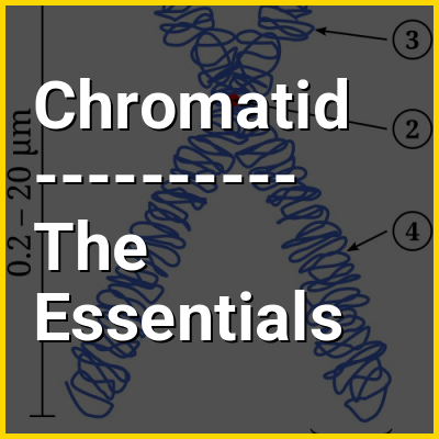
A chromatid (Greek khrōmat- 'color' + -id) is one half of a duplicated chromosome. Before replication, one chromosome is composed of one DNA molecule. In replication, the DNA molecule is copied, and the two molecules are known as chromatids. During the later stages of cell division these chromatids separate longitudinally to become individual chromosomes.

Chromatid pairs are normally genetically identical, and said to be homozygous. However, if mutations occur, they will present slight differences, in which case they are heterozygous. The pairing of chromatids should not be confused with the ploidy of an organism, which is the number of homologous versions of a chromosome.

↓ Menu

>>>PUT SHARE BUTTONS HERE<<<
In this Dossier

Chromatid in the context of Karyotype

A karyotype is the general appearance of the complete set of chromosomes in the cells of a species or in an individual organism, mainly including their sizes, numbers, and shapes. Karyotyping is the process by which a karyotype is discerned by determining the chromosome complement of an individual, including the number of chromosomes and any abnormalities.

A karyogram or idiogram is a graphical depiction of a karyotype, wherein chromosomes are generally organized in pairs, ordered by size and position of centromere for chromosomes of the same size. Karyotyping generally combines light microscopy and photography in the metaphase of the cell cycle, and results in a photomicrographic (or simply micrographic) karyogram. In contrast, a schematic karyogram is a designed graphic representation of a karyotype. In schematic karyograms, just one of the sister chromatids of each chromosome is generally shown for brevity, and in reality they are generally so close together that they look as one on photomicrographs as well unless the resolution is high enough to distinguish them. The study of whole sets of chromosomes is sometimes known as karyology.

↑ Return to Menu

Chromatid in the context of Chromosome

A chromosome is a package of DNA containing part or all of the genetic material of an organism. In most chromosomes, the very long thin DNA fibers are coated with nucleosome-forming packaging proteins; in eukaryotic cells, the most important of these proteins are the histones. Aided by chaperone proteins, the histones bind to and condense the DNA molecule to maintain its integrity. These eukaryotic chromosomes display a complex three-dimensional structure that has a significant role in transcriptional regulation.

Normally, chromosomes are visible under a light microscope only during the metaphase of cell division, where all chromosomes are aligned in the center of the cell in their condensed form. Before this stage occurs, each chromosome is duplicated (S phase), and the two copies are joined by a centromere—resulting in either an X-shaped structure if the centromere is located equatorially, or a two-armed structure if the centromere is located distally; the joined copies are called 'sister chromatids'. During metaphase, the duplicated structure (called a 'metaphase chromosome') is highly condensed and thus easiest to distinguish and study. In animal cells, chromosomes reach their highest compaction level in anaphase during chromosome segregation.

↑ Return to Menu

Chromatid in the context of Chromosomal crossover

Chromosomal crossover, or crossing over, is the exchange of genetic material during sexual reproduction between two homologous chromosomes' non-sister chromatids that results in recombinant chromosomes. It is one of the final phases of genetic recombination, which occurs in the pachytene stage of prophase I of meiosis during a process called synapsis. Synapsis is usually initiated before the synaptonemal complex develops and is not completed until near the end of prophase I. Crossover usually occurs when matching regions on matching chromosomes break and then reconnect to the other chromosome, resulting in chiasma which are the visible evidence of crossing over.

↑ Return to Menu

Chromatid in the context of Cytokinesis

Cytokinesis (/ˌstkɪˈnsɪs/) is the part of the cell division process and part of mitosis during which the cytoplasm of a single eukaryotic cell divides into two daughter cells. Cytoplasmic division begins during or after the late stages of nuclear division in mitosis and meiosis. During cytokinesis the spindle apparatus partitions and transports duplicated chromatids into the cytoplasm of the separating daughter cells. It thereby ensures that chromosome number and complement are maintained from one generation to the next and that, except in special cases, the daughter cells will be functional copies of the parent cell. After the completion of the telophase and cytokinesis, each daughter cell enters the interphase of the cell cycle.

Particular functions demand various deviations from the process of symmetrical cytokinesis; for example, in oogenesis in animals, the ovum takes almost all the cytoplasm and organelles. This leaves very little for the resulting polar bodies, which in most species die without function, though they do take on various special functions in other species.Another form of mitosis occurs in tissues such as liver and skeletal muscle; it omits cytokinesis, thereby yielding multinucleate cells (see syncytium).

↑ Return to Menu

Chromatid in the context of Sister chromatid

A sister chromatid refers to the identical copies (chromatids) formed by the DNA replication of a chromosome, with both copies joined together by a common centromere. In other words, a sister chromatid may also be said to be 'one-half' of the duplicated chromosome. A pair of sister chromatids is called a dyad. A full set of sister chromatids is created during the synthesis (S) phase of interphase, when all the chromosomes in a cell are replicated. The two sister chromatids are separated from each other into two different cells during mitosis or during the second division of meiosis.

Compare sister chromatids to homologous chromosomes, which are the two different copies of a chromosome that diploid organisms (like humans) inherit, one from each parent. Sister chromatids are by and large identical (since they carry the same alleles, also called variants or versions, of genes) because they derive from one original chromosome. An exception is towards the end of meiosis, after crossing over has occurred, because sections of each sister chromatid may have been exchanged with corresponding sections of the homologous chromatids with which they are paired during meiosis. Homologous chromosomes might or might not be the same as each other because they derive from different parents.

↑ Return to Menu

Chromatid in the context of Telophase

Telophase (from Ancient Greek τέλος (télos) 'end, result, completion' and φάσις (phásis) 'appearance') is the final stage in both meiosis and mitosis in a eukaryotic cell. During telophase, the effects of prophase and prometaphase (the nucleolus and nuclear membrane disintegrating) are reversed. As chromosomes reach the cell poles, a nuclear envelope is re-assembled around each set of chromatids, the nucleoli reappear, and chromosomes begin to decondense back into the expanded chromatin that is present during interphase. The mitotic spindle is disassembled and remaining spindle microtubules are depolymerized. Telophase accounts for approximately 2% of the cell cycle's duration.

Cytokinesis typically begins before late telophase and, when complete, segregates the two daughter nuclei between a pair of separate daughter cells.

↑ Return to Menu

Chromatid in the context of Locus (genetics)

In genetics, a locus (pl.: loci) is a specific, fixed position on a chromosome where a particular gene or genetic marker is located. Each chromosome carries many genes, with each gene occupying a different position or locus; in humans, the total number of protein-coding genes in a complete haploid set of 23 chromosomes is estimated at 19,000–20,000.

Genes may possess multiple variants known as alleles, and an allele may also be said to reside at a particular locus. Diploid and polyploid cells whose chromosomes have the same allele at a given locus are called homozygous with respect to that locus, while those that have different alleles at a given locus are called heterozygous. The ordered list of loci known for a particular genome is called a gene map. Gene mapping is the process of determining the specific locus or loci responsible for producing a particular phenotype or biological trait. Association mapping, also known as "linkage disequilibrium mapping", is a method of mapping quantitative trait loci (QTLs) that takes advantage of historic linkage disequilibrium to link phenotypes (observable characteristics) to genotypes (the genetic constitution of organisms), uncovering genetic associations.

↑ Return to Menu

Chromatid in the context of Spindle checkpoint

The spindle checkpoint, also known as the metaphase-to-anaphase transition, the spindle assembly checkpoint (SAC), the metaphase checkpoint, or the mitotic checkpoint, is a cell cycle checkpoint during metaphase of mitosis or meiosis that prevents the separation of the duplicated chromosomes (anaphase) until each chromosome is properly attached to the spindle. To achieve proper segregation, the two kinetochores on the sister chromatids must be attached to opposite spindle poles (bipolar orientation). Only this pattern of attachment will ensure that each daughter cell receives one copy of the chromosome. The defining biochemical feature of this checkpoint is the stimulation of the anaphase-promoting complex by M-phase cyclin-CDK complexes, which in turn causes the proteolytic destruction of cyclins and proteins that hold the sister chromatids together.

↑ Return to Menu