The thoracic wall or chest wall is the boundary of the thoracic cavity.
HINT:
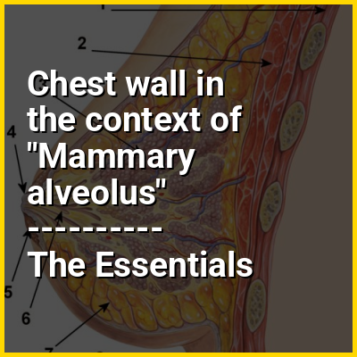
👉 Chest wall in the context of Mammary alveolus
A mammary alveolus (pl.: alveoli, from Latin alveolus, "little cavity") is a small cavity or sac found in the mammary gland. Mammary alveoli are the site of milk production and storage in the mammary gland. Mammary alveoli cluster into groups called mammary lobules, and each breast may contain 15 to 20 of these lobules. The lobules drain milk through the lactiferous ducts out of the nipples.
In this Dossier
- ⭐ Core Definition: Chest wall
- 👉 Chest wall in the context of Mammary alveolus
- Chest wall in the context of Pleurae
- Chest wall in the context of Pneumothorax
- Chest wall in the context of Intercostal muscle
Chest wall in the context of Pleurae
The pleurae (sg.: pleura) are the two flattened pleural sacs filled with pleural fluid that surround each lung, and lines their surrounding tissues. They are formed of two opposing layers of serous membrane that separate the lungs from the mediastinum, the inside surfaces of the surrounding chest walls and the diaphragm. Although wrapped onto itself resulting in a double layer, each lung is surrounded by a single, continuous pleural membrane.
The pleura that covers the surface of each lung is the visceral pleura. The pleura typically dips between the lobes of the lung as fissures, and is formed by the invagination of lung buds into each thoracic sac during embryonic development. The pleura that lines the chest wall, is the parietal pleura.
View the full Wikipedia page for PleuraeChest wall in the context of Pneumothorax
A pneumothorax is collection of air in the pleural space between the lung and the chest wall. Symptoms typically include sudden onset of sharp, one-sided chest pain and shortness of breath. In a minority of cases, a one-way valve is formed by an area of damaged tissue, in which case the air pressure in the space between chest wall and lungs can be higher; this has been historically referred to as a tension pneumothorax, although its existence among spontaneous episodes is a matter of debate. This can cause a steadily worsening oxygen shortage and low blood pressure. This could lead to a type of shock called obstructive shock, which could be fatal unless reversed. Very rarely, both lungs may be affected by a pneumothorax. It is often called a "collapsed lung", although that term may also refer to atelectasis.
A primary spontaneous pneumothorax is one that occurs without an apparent cause and in the absence of significant lung disease. Its occurrence is fundamentally a nuisance. A secondary spontaneous pneumothorax occurs in the presence of existing lung disease. Smoking increases the risk of primary spontaneous pneumothorax, while the main underlying causes for secondary pneumothorax are COPD, asthma, and tuberculosis. A traumatic pneumothorax can develop from physical trauma to the chest (including a blast injury) or from a complication of a healthcare intervention.
View the full Wikipedia page for PneumothoraxChest wall in the context of Intercostal muscle
The intercostal muscles comprise many different groups of muscles that run between the ribs, and help form and move the chest wall. The intercostal muscles are mainly involved in the mechanical aspect of breathing by helping expand and shrink the size of the chest cavity.
View the full Wikipedia page for Intercostal muscle